|
Finding disease-causing gene mutations is seen
as the shortest cut to preventing or curing heritable
diseases. However, each such procedure is resource-consuming
and costly, and it becomes especially cost-ineffective for
rare hereditary diseases. In this story, Edwin M. Stone,
M.D., Ph.D., director of the Carver Laboratory for
Molecular Diagnosis, tells how his group came up with a
cost-effective solution to genetic testing procedures for
hereditary eye diseases. |
History: Inherited diseases are caused by variations in
DNA. Therefore, it is theoretically possible to diagnose any genetic
disease by examining a patient's DNA for such variations. However,
until very recently, a number of technical, financial, and ethical
reasons have kept such testing out of the medical mainstream. Before
the advent of DNA cloning in the 1970s, the only way to detect any
sequence variation in the DNA of higher organisms was to observe
variations in the proteins they encoded. This method allowed
researchers to deduce the molecular nature of some genetic diseases,
such as sickle-cell anemia. However, the great majority of
disease-causing variations remained buried in the vast featureless
and colorless polymer that is the human genome.
In the 1960s and 1970s, researchers discovered restriction
enzymes and learned to use them to insert small bits of exogenous
DNA into plasmids and viruses for replication. This process allowed
individual genes to be isolated for the first time. It also made it
possible to identify sequence variations within the human genome
even when such variations did not cause a detectable change in an
accessible protein. However, it was the development of the
polymerase chain reaction (PCR) in the 1980s that made it possible
to perform the types of genetic testing that are available today.
PCR has made it possible to perform tests on DNA samples
consisting of only nanograms of DNA, while previous methods required
more than a thousand times this amount. PCR also lends itself to
automation and speed so that a single technician can perform an
experiment to evaluate a single genetic locus in 400 or 500 people
in a single day. In the past decade, the combination of cloning,
PCR, automated DNA sequencing, and computer analysis of the
resulting sequences has made it possible to embark upon the sequence
analysis of entire genomes (e.g., the Human Genome Project); the
genomic sequences of several model organisms, including humans, have
now been completed.
When specific human genes are screened for variations in a large
number of different individuals, a large amount of variations is
commonly found that does not appear to alter the structure and
function of an individual. These silent variations are so common
that any two human beings will differ from one another at least once
every thousand nucleotides. This fact, coupled with the knowledge
that the haploid human genome consists of more than three billion
nucleotides, means that any two normal individuals will differ from
one another at at least three million positions. Thus, finding a
single nucleotide variation that is responsible for human disease is
like trying to find a single steel needle in a haystack full of
three million silver needles.
New facts: True disease-causing mutations can be
distinguished from nondisease-causing polymorphisms in a number of
ways, but the most common is to correlate the inheritance of a
disease within a large family with the inheritance of a specific
molecular variation. The second most common way is to use a similar
correlative approach to evaluate a large number of unrelated
individuals affected with a given disease and to demonstrate that a
variation within a candidate gene is found more often among
individuals with the disease than in ethnically similar unaffected
individuals. Once such a correlation has been established in a
statistically rigorous way, this information becomes the basis of a
clinical molecular test. One might imagine that at this point, a
for-profit testing firm could use such information to develop a
commercially viable test and that in a fairly short period of time
this test would be widely available to the medical community.
Although this has happened in some cases, many inherited
disorders are so rare and the number of different disease-causing
variations so numerous that there is simply insufficient demand to
make such a test commercially viable. This is complicated by the
fact that many diagnostically useful genomic sequences have been
patented and that the legal costs involved in obtaining the rights
to use the necessary intellectual property have made marginally
viable genetic tests impossible to sustain.
For all of these reasons, most genetic testing for rare human
diseases in the past decade has been performed on a research basis
in the same laboratories that actually discovered the
disease-causing genes. Although this was a reasonable approach for
the first few years of human gene discovery, the past decade's
explosive increase in the knowledge of disease-causing variations
quickly exhausted the ability of research laboratories to keep pace.
In response to this challenge, University of Iowa Health Care
researchers have explored a new strategy for providing practical
genetic testing for rare eye diseases for the entire U.S. This
strategy involves a partnership between the University of Iowa Roy
J. and Lucille A. Carver College of Medicine, UI Hospitals and
Clinics, the Department of Pathology, the research laboratories of
the Center for Macular Degeneration, and the owners of the
intellectual property of the relevant genes. In this approach,
academic physicians use their expertise to design, perform, and
interpret the tests and charge the patient only for the costs that
are actually incurred in performing them (e.g., reagents and
technician time).
In most cases to date, gene sequence patent holders have allowed
their intellectual property to be used at no cost for this nonprofit
endeavor, while in a few cases, they have asked for only a modest
fee per test. The creation of the non-profit genetic testing
laboratory at UI Hospitals and Clinics was greatly aided by a
multi-million dollar endowment that was jointly provided by the Roy
J. Carver Charitable Trust and the Department of Ophthalmology and
Visual Sciences. For this reason, the laboratory is now known as the
Carver Laboratory for Molecular Diagnosis.
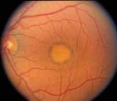
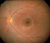
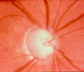
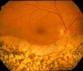
Practice: The goal of the Carver Laboratory is to provide
a wide variety of clinically useful tests, a rapid turnaround time,
and an easily interpreted written report--all at a modest cost. Most
tests offered by the laboratory cost well under $500. The most
expensive test offered, involving the analysis of six different
genes, costs a little over $1,000 (Table
1). In addition, the Center for Macular Degeneration offers
expert genetic counseling regarding the tests' results for patients
who do not have access to such counseling in their home communities.
The Carver Laboratory maintains a web page (http://www.carverlab.org/) with
a wide array of information for physicians and patients about
inherited eye diseases and eye disease genes. The web page can be
used to learn more about the clinical features of these diseases,
their mode of inheritance, and the ways in which patients can
benefit from genetic testing. Visitors to the web page also have
access to a variety of data about these genes, which have been
collected by research laboratories at the UI Carver College of
Medicine over the past 15 years.
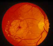
The Molecular Ophthalmology Laboratory has screened thousands of
patients with inherited eye diseases for variations in a large
number of disease-associated genes. The knowledge gained from this
experience is essential to the development of genetic tests that
will benefit the patient. For example, some genes are very large
while others are quite small. For large genes, it is not very
efficient to screen the entire gene for variations, because most
large genes have regions that harbor few if any disease-causing
changes.
It is much more efficient to stratify the screening so that the
regions of a gene that are known to harbor the most variations are
screened first, with less promising regions screened later or not at
all. For diseases that are caused by several different genes, such
as Leber Congenital Amaurosis (LCA), the genes are not screened
individually. Instead, the subparts of all six LCA genes are ranked
according to the likelihood of harboring a disease-causing change,
and these regions are screened in the order of decreasing likelihood
of a significant finding.
LCA is a recessive disease, and affected patients are expected to
carry two mutations--one inherited from each parent. Thus, once the
first disease-causing variation is identified with the probability
stratification approach described above, attention can then be
shifted to finding the second mutation in the same gene. This
sequential screening strategy, which takes full advantage of the
large body of research data collected at the College in the past,
can detect meaningful disease-causing mutations much faster and at a
much lower cost than more conventional approaches.
The Carver Laboratory website features a table that summarizes
the genomic structures of 34 genes that contribute to 19 different
human eye diseases, as well as the laboratory's cumulative screening
experience with these genes. As new variations are detected by the
nonprofit testing service, these changes will also be added to the
web-based database.
|
|